Color-coded longitudinal interventricular septal tissue velocity imaging, strain and strain rate in healthy Doberman Pinschers

Author information
Simak J., Keller L., Killich M., Hartmann K., Wess G. Color-coded longitudinal interventricular septal tissue velocity imaging, strain and strain rate in healthy Doberman Pinschers // J Vet Cardiol. 2011 Mar;13(1):1-11.
Abstract
OBJECTIVE: To determine reference values for septal longitudinal tissue Doppler imaging (TDI) in Doberman Pinschers.
BACKGROUND: TDI includes several new techniques, such as tissue velocity imaging (TVI), strain, and strain rate that might be used to detect early myocardial dysfunction. However, before these techniques can be used, breed specific reference ranges need to be established.
ANIMALS, MATERIALS AND METHODS: One-hundred healthy Doberman Pinschers ≥ 4 years of age were prospectively evaluated using electrocardiography, Holter recording, and echocardiography. Systolic, early and late diastolic parameters of septal longitudinal color-coded TVI, strain, and strain rate were analyzed in three myocardial segments (basal, middle, and apical).
RESULTS: TDI was feasible in all Doberman Pinschers. Reference values were established for every myocardial segment. There was a significant velocity gradient from the basal to the apical segment for all TVI parameters. No differences between the three segments were found for systolic strain and strain rate. Several diastolic strain rate values were significantly different between myocardial segments. Influence of age, weight, and gender was not clinically relevant.
CONCLUSIONS: This study provides reference values for TVI, strain, and strain rate in Doberman Pinschers. The parameters established here can be used for a supportive diagnostic approach.
Introduction
Dilated cardiomyopathy (DCM) is the most common acquired cardiac disease in large breed dogs. Doberman Pinschers have a particularly high prevalence for this disease.1-3,a-c In a family of Doberman Pinschers the disease was inherited as an autosomal dominant trait.4 A typical feature of DCM in this breed is the disease progression in several stages. Disturbances at the cellular level of the myocardium, which are not detectable by routinely applied diagnostic methods, are followed by the occult stage, during which patients do not show any clinical signs. Ventricular tachyarrhythmias are commonly seen in the occult stage, whereas echocardiographic changes may or may not occur at this stage.2,5 Syncope caused by ventricular tachyarrhythmias or sudden cardiac death can occurin the occult phase of the disease. In the later course of the disease dogs develop apparent systolic myocardial dysfunction, which can be detected by echocardiography. As the disease progresses, dogs develop congestive heart failure and overt clinical symptoms.2,5-7 Cardiomyopathy in Doberman Pinschers in the occult stage of the disease can be detected by 24-h ambulatory electrocardiography (Holter) or by conventional echocardiography.2,8 Currently the disease can only be diagnosed, once the dogs have electrocardiographic changes detectable using Holter, or once the dogs have changes detectable by conventional echocardiography. However, changes on a cellular basis, that lead to myocardial dysfunction, but not yet systolic or diastolic chamber dilation or global systolic dysfunction cannot be demonstrated. For such an early diagnosis more sensitive methods would be necessary.
Tissue Doppler imaging (TDI) includes several new techniques, such as tissue velocity (TVI), strain, and strain rate imaging. These new, sensitive, and non-invasive techniques are increasingly used for the evaluation of systolic and diastolic myocardial function in dogs.9-14 TVI was the first TDI method established and it is used in the majority of studies in dogs.11,12,15-18 However, TVI is not able to differentiate between active and passive motion of myocardial segments. In humans, the newer techniques strain and strain rate are less influenced by overall cardiac motion and tethering effects in adjacent segments than TVI and therefore overcome the limitations of TVI. They appear to be sensitive indicators of myocardial diseases.19,20 However, only few studies exist concerning strain and strain rate imaging in dogs.9,10,14
Several case reports and clinical studies evalu- atingTVI, showed that it is a promising technique for the detection of early myocardial dysfunction in preclinical stages of dogs with cardiomyopathy. TVI measurements in these dogs were abnormal, while conventional echocardiography was still unremarkable and only became abnormal at a later stage.16-18 One study in Doberman Pinschers with occult or overt DCM demonstrated reduced systolic and diastolic pulsed-wave TVI mitral annular velocities.21 One single report evaluated systolic and diastolic TVI and strain parameters in canine patients of different breeds with spontaneous DCM.10 Measuring interventricular septal longitudinal motion is a standard method in humans to evaluate myocardial func- tion.22,23 Longitudinally directed fibers compose only a small portion of the myocardial mass, but several studies have shown that the systolic motion of the atrioventricular plane toward the apex is an important component of the systolic performance of the left ventricle and quantification of this movement amplitude will provide important information about left ventricular systolic function.24-26
Repeatability and reproducibiltyofTDI in healthy awake dogs is acceptable.9,12,14 An influence of breed on TDI values has been reported and breed specific reference ranges were recommended.11,14 Additionally, TDI may be influenced by age, body weight and sex, but these factors were not investigated in a homogenous population of dogs.11 In summary, TDI has not been evaluated in a large group of Doberman Pinschers. However, before these methods can be used in clinical practice, normal values need to be established. The aim of this study was therefore toestablish reference ranges for color-coded interventricular septal longitudinal TVI, strain and strain rate in healthy Doberman Pinschers. In addition, the influence of the physiologic parameters age, body weight, and sex on TDI parameters was evaluated and TVI, strain, and strain rate in different myocardial segments (basal, middle, and apical) were compared.
Animals, materials and methods Study population
This prospective study included 100 client-owned healthy Doberman Pinschers > 4 years of age. Written owner consent was obtained before inclusion in the study. Dogs were presented to the Clinic of Small Animal Medicine, Ludwig-Maximilian- University, Munich, Germany for screening purposes for cardiomyopathy between August 2004 and July 2009. Participating dogs had to be > 4 years of age. An unremarkable history, physical examination, electrocardiogram (ECG), 24-h Holter recording, and conventional echocardiography confirmed that the dogs were healthy. This study was in accordance with the German Animal Welfare Act.
Inclusion criteria: Criteria to include the dogs as healthy were a normal 5-min ECGd (sinus rhythm or respiratory sinus arrhythmia) without any premature complexes. Holter recordingse with < 50 ventricular premature complexes (VPCs)/24-h were considered normal. Left ventricular internal dimensions at end systole (LVIDs) and end diastole (LVIDd) had to be within established reference ranges for Doberman Pinschers (LVIDd < 48.0 mm, LVIDs < 38.0 mm) and left atrium to aorta ratio (LA/Ao) had to be < 1.5.27-29 Exclusion criteria: Dogs with evidence of cardiac or systemic diseases (detected by history, physical examination, ECG, 24-h Holter recording, or conventional echocardiography) were excluded. None of the dogs received any cardiac or other drugs.
Echocardiography and tissue Doppler imaging analysis
Echocardiographyf was performed by three experienced investigators (LK, MK, and GW) in unsedated dogs positioned in right and left lateral recumbency with a simultaneous ECG tracing. A 2.0/3.5 MHz transducer was used for echocardiographic examination. To exclude cardiac disease, conventional two-dimensional echocardiography, color flow, continuous wave, and pulsed-wave Doppler examinations were assessed in every dog. M-mode measurements had to be within the reported reference ranges.27-30 Velocities over the aortic and pulmonary valves were measured using continuous wave Doppler examinations and had to be below 2.2 m/s. Each variable was measured at least three times and the average values were used. Echocardiography was performed according to the recommendations of the Echocardiography Committee of the Specialty of Cardiology, American College of Veterinary Internal Medicine.31
For TDI analysis the interventricular septum (IVS) was recorded as a single wall from a standard left apical four chamber view parallel to the ultrasound beam to avoid angular errors. Grayscale images were superimposed by color tissue Doppler. The TDI frame rate had to exceed 180 frames/s. Loops showing aliasing were excluded from the analysis. Three consecutive cardiac cycles were stored digitally for further offline analysis.
TDI data were analyzed by one experienced observer (JS) using a commercial software pack- age.g Septal endocardial borders were manually traced and semi-automatic tracking of the myocardial wall was performed by the software. This ensured, that the software followed the myocardial wall throughout the cardiac cycle. The two-dimensional grayscale image was only used for semi-automatic tracking, but color-Doppler data were used for further analysis. The IVS was divided by the software automatically into three myocardial segments (basal, middle, and apical). Color Doppler derived systolic (S), early diastolic (E), and late diastolic (A) peaks using TVI (cm/s) and strain rate (s-1) methods and peak systolic strain (%) were measured in each myocardial segment of the IVS. The software automatically averaged out several single point measurements to one average value over the whole myocardial segment.
Six echocardiograms were randomly selected to be subjected to three repeated analyses by the same investigator (JS) to determine intrareader measurement variability (within-day and between- day) for all TDI data analyzed as described above. The same six echocardiograms were subjected to independent repeated analyses by a second investigator (GW) to determine interreader measurement variability. Both investigators were unaware of the results of the prior echocardio- graphic analyses. The within- and between-day variations of the involved echocardiographers were also tested. Ultrasound examinations of six Doberman Pinschers were acquired by all of the three echocardiographers (LK, MK, and GW) twice in one day to calculate the intraechocardiographer within-day variability. To determine the between- day intraechocardiographer variability, the echo- cardiographic examination of the six Doberman Pinschers was repeated by each of the three echocardiographers within 10 days. The resultant echocardiograms were analyzed by the same investigator (JS).
Statistical analysis
Statistical analysis was performed by commercially available statistical software.h Descriptive statistics (mean, standard deviation, median, and range) and frequencies were calculated for physiologic (age, sex, or weight), conventional echo- cardiographic, and TDI parameters. Reference values were calculated as mean ± the standard deviation (SD). Normal distribution of the data was evaluated by histograms, Q-Q-plots and a Kolmogorov—Smirnov analysis. One-way analysis of variance was used to investigate differences between myocardial segments. Bonferroni analysis was used as a posthoc test when analysis of variance showed a significant difference between the groups. The results were illustrated as boxplots. Influence of age, gender, and body weight on TDI values was tested by multivariate analysis of variance and the significant differences were displayed as boxplots or scatterplots. The intra- and interobserver coefficients of variation (CV) were calculated using a variance component analysis. The CVs were obtained by dividing the root of the variance error by the mean of the repeated measurements, times 100. Significance was defined as P < 0.05.
Results
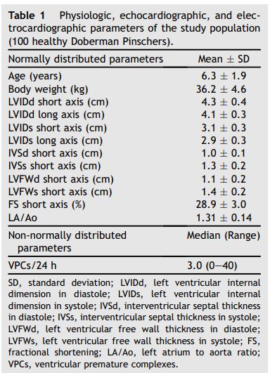
A total of 100 Doberman Pinschers (44 male and 56 female dogs) were classified as healthy according to the inclusion criteria. Physiologic parameters (age and body weight), conventional echocardio- graphic data (LVIDd, LVIDs, IVS in diastole and systole, left ventricular free wall in diastole and systole, fractional shortening, and LA/Ao) and number of VPCs are displayed in Table 1.
Septal longitudinal TDI was feasible with good image quality in all 100 Doberman Pinschers. All dogs showed positive S and negative E and A waves in all myocardial segments using TVI evaluation. Systolic peaks for strain and systolic strain rate were negative in all dogs, whereas diastolic strain rate waves (E and A) were positive in every Doberman Pinscher. Reference values for TVI, strain, and strain rate were calculated as averages for each myocardial segment (basal, middle, and apical) and displayed as mean values ± SD (Table 2). Concerning TVI and strain rate, isovolumic contraction and isovolumic relaxation waves were always present between the end of the A wave and the beginning of the S wave and the end of the S wave and the beginning of the E wave, respectively (Fig. 1).
Table 1 Physiologic, echocardiographic, and electrocardiographic parameters of the study population (100 healthy Doberman Pinschers)
Comparing septal longitudinal systolic and diastolic TVI parameters between the three myocardial segments, a significant velocity gradient from the basal to the apical segment was found for all velocities measured, with the highest values in the basal segment (P < 0.001). There was no significant difference between the segments concerning strain and systolic strain rate values. Diastolic strain rate showed significant differences between the apical and midventricular E wave, the apical and basal E wave, and the apical and basal A wave (P < 0.001), showing a subtle gradient with the highest values in the apical segment (Fig. 2).
Overall there was almost no influence of age, sex, and body weight on septal longitudinal TDI parameters. Only strain rate A in the apical segment was influenced by age with significantly higher values in older dogs (P = 0.005). TVI and strain rate E waves in the basal segment were influenced by sex, with higher values in males compared to female dogs (P < 0.05). Boxplots and scatterplots for the significant physiological influencing factors are shown in Fig. 3.
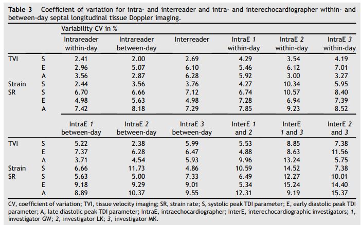
Results of the repeatability and reproducibility study were determined (Table 3). All CV values were < (range 2.00—15.37).
Table 2 Septal longitudinal tissue Doppler imaging parameters of different myocardial segments (basal, middle, and apical) of 100 healthy Doberman Pinschers

Figure 1 Examples of septal longitudinal tissue Doppler imaging curves of a healthy Doberman Pinscher obtained from left apical four chamber view
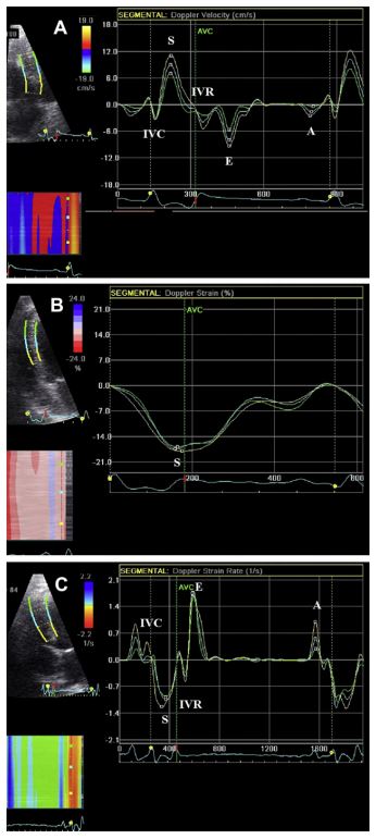
A, late diastolic wave; AVC, aortic valve closure; E, early diastolic wave; IVC, isovolumic contraction time; IVR, iso- volumic relaxation time; S, systolic wave; blue, middle segment of interventricular septum; green, apical segment of interventricular septum; yellow, basal segment of interventricular septum. A: tissue velocity imaging curves (cm/s). B: strain curves (%). C: strain rate curves (s~1).
Figure 2 Boxplots of peak tissue Doppler imaging values in different interventricular septal myocardial segments, measured in 100 healthy Doberman Pinschers
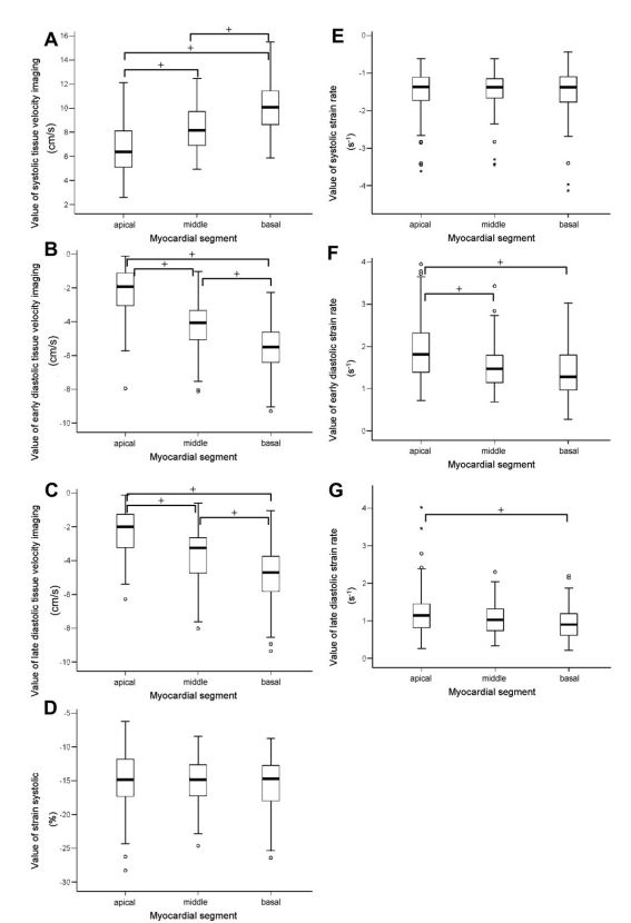
A: systolic tissue velocity imaging (cm/s). B: early diastolic tissue velocity imaging (cm/s). C: late diastolic tissue velocity imaging (cm/s). D: systolic strain (%). E: systolic strain rate (s~1). F: early diastolic strain rate (s~1). G: late diastolic strain rate (s~1). +, P < 0.05; o, outlier; *, extreme value.
Figure 3 Scatterplots (A) and boxplots (B and C) in 100 healthy Doberman Pinschers showing distribution of statistically significant tissue Doppler imaging parameters influenced by age or sex
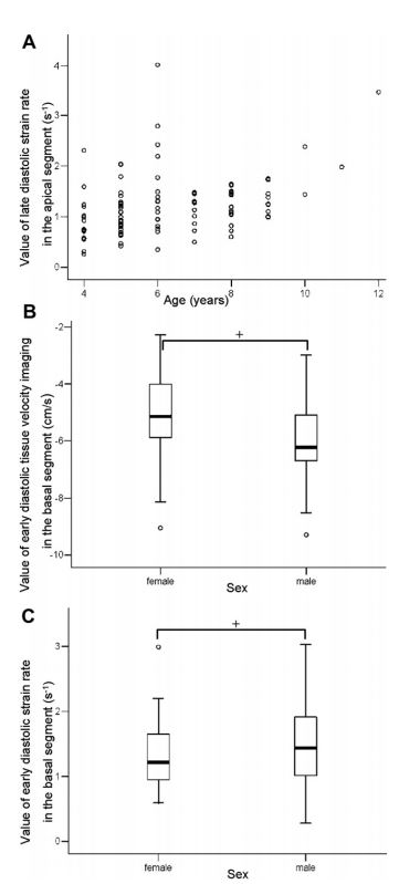
A: late diastolic strain rate in the apical segment and age; P = 0.005. B: early diastolic tissue velocity in the basal segment and sex. C: early diastolic strain rate in the basal segment and sex. +, P < 0.05; o, outlier (only in Fig. B and C).
Discussion
The reference values for septal longitudinal TVI, strain and strain rate, established in this prospective study, set the basis for the use of these new methods in the diagnostic approach to cardiomyopathy in Doberman Pinschers. Unlike other breeds, Doberman Pinschers have a deep narrow chest, which causes an oblong heart shape and therefore often a subjectively decreased systolic function, compared to other breeds. This different heart shape, compared to other breeds, may affect the motion of the heart. A previous study found significant differences between TVI values of different breeds and as a consequence it has been recommended to establish breed specific reference ranges for color-coded TVI methods.11 Reference ranges for color-coded TVI, strain and strain rate measurements are a prerequisite to use these new methods in clinical practice. However, no reference values existed for color-coded TDI methods for Doberman Pinschers, a breed in which TDI might be especially useful due to the high prevalence of DCM in this breed. Septal longitudinal TDI measurements were feasible with good quality in all 100 dogs participating in this study. Therefore septal longitudinal colorTDI represents an almost universally applicable method in this breed. To date only few reports exist about longitudinal interventricular septal myocardial motion evaluated by colorTDI in dogs.9,14,32 All Doberman Pinschers in this study showed normal waveformswith positive S and negative E andAwaves for TVI, negative S and positive E and A strain rate waves, and a negative S strain in accordance with other studies in dogs and humans.9,11,12,33-35 This finding indicates that inverse waves should be considered pathologic. Isovolumic contraction or isovolumic relaxation waves were present between the end of the A wave and the beginning of the S wave and the end of the S wave and the beginning of the A wave, respectively.11,12 TDI might be therefore an alternative method to measure isovolumic contraction and relaxation times and those values might be less influenced by changes in preload and afterload compared to conventional echocardiographic measurements.36,37
In the present study peak S septal longitudinal strain rate and strain in Doberman Pinschers were lower than values reported in a study examining dogs of different breeds.9 This fact confirms the subjective impression of a reduced systolic function even in healthy Doberman Pinschers, compared to other breeds. This impaired systolic function of healthy Doberman Pinschers (compared to other breeds) was first documented by conventional echocardiography and cardiac catheterization.38 To our knowledge currently only one other study exists evaluating systolic and diastolic color TDI in the IVS in dogs.14 In another report, only systolic strain and strain rate in the IVS were evaluated.9
Table 3 Coefficient of variation for intra- and interreader and intra- and interechocardiographer within- and between-day septal longitudinal tissue Doppler imaging
Using the IVS instead of the LVFW has several potential advantages. In our experience obtaining image planes parallel to the ultrasound beam is easier for the IVS, compared to the LVFW. Potentially it could be more difficult to align with the IVS or LVFW of a Doberman Pinscher with DCM and a rounded left ventricle than with the same structures ina healthy dog with an elliptical left ventricle. Further studies are required to evaluate TDI in Doberman Pinschers with DCM. Breathing artifacts due to lung tissue overlaying the image plane and reverberation artifacts are more often a problem using the LVFW than using the IVS. Peak systolic TVI in the LVFW of other breeds was slightly higher compared to the values reported in the present study using the IVS in Doberman Pinschers. Diastolic left ventricular velocities vary in other breeds.11,12 These observed differences might be a consequence of the other studies examining dogs of different breeds, or examining different myocardial walls (IVS versus LVFW). The subjective impression and previously documented evidence of decreased systolic function in healthy Doberman Pinschers, was believed to be a consequence of apparently decreased short axis contractility brought about by the oblong shape of the left ventricle in this breed. It was presumed that there would be normal apico- basilar function. However, the results of this study also show reduced longitudinal myocardial function. It would be of interest to compare these findings with radial and circumferential motion measured by TDI in healthy Doberman Pinschers. Additionally, the IVS can be influenced by right ventricular function. However, in Doberman Pinschers with suspicion of DCM an impression of global myocardial performance including the right ventricle could be helpful.
To date only one study has evaluated TVI in Doberman Pinschers with different stages of DCM. In that study real-time pulsed-wave TVI of the mitral annulus was used.21 Healthy Doberman Pinschers showed higher systolic and diastolic velocities than dogs with occult or overt DCM. The pulsed-wave TVI data of the control group were clearly higher than the results of the present study using color-Doppler TVI. The finding that pulsed- wave TVI methods reveal significantly higher values, compared to color-Doppler TVI has been reported in humans and dogs.39,i This also shows, that reference ranges established by pulsed-wave Doppler TVI cannot be used interchangeably with color-coded TVI methods, even if the same breeds
Comparing septal longitudinal systolic and diastolic TVI parameters between the three myocardial segments, a statistically significant velocity gradient from the basal to the apical segment was found for all velocities (S, E, and A) measured, with the highest values in the basal segment (P < 0.001). This finding is in accordance with recent studies in human and veterinary medicine.11,39,40 In contrast to strain and strain rate imaging, TVI is influenced by overall cardiac motion and contraction in adjacent myocardial segments. In accordance with other studies systolic strain and strain rate values were distributed more homogeneously between the myocardial segments.40,41 In contrast to this study, another veterinary study reported significantly higher systolic strain and strain rate values in the basal segment compared to the apical myocardial segment, but these data were derived from a much smaller population than is reported in the current study.9 Only diastolic strain rate values were different between the apical and the other myocardial segments for E and A waves. Because of the homogenous distribution of systolic strain and strain rate parameters, values can be interpreted independently of the myocardial segment used for analysis. Due to these results it could be useful for clinical applications to combine the systolic strain or strain rate values of different myocardial segments to one global septal parameter to evaluate overall myocardial performance. In contrast, for TVI and diastolic strain rate individual reference ranges for each specific myocardial segment should be used. Furthermore, in cases of regional myocardial dysfunction assessing isolated myocardial segments could be useful.
In the present study septal longitudinal TDI parameters were only partially influenced by physiological parameters. Concerning age, only strain rate E in the apical myocardial segment showed higher values with increasing age. This finding is similar to other veterinary and human reports where no correlation between age and longitudinal strain rate was found. However, it might be possible that the study failed to identify an influence of age on most diastolic parameters, because the mean age (6.3 years) was too low for an age-dependent diastolic impairment, even though dogs up to twelve years were included.
Regarding gender, male dogs had significantly higher values of TVI and strain rate E waves in the basal segment. All other results were not influenced by sex. This is in accordance to a study in humans, where no influence of sex on strain rate was found.9,25 As the physiological influence factors found in this study were only mild and significantly different in only a few myocardial segments, the influence of these factors is in our opinion not clinically relevant in Doberman Pinschers. In summary, TVI, strain and strain rate are only mildly influenced by age and sex and not influenced by body weight in Doberman Pinschers. This could be an advantage over other echocardiographic parameters.
The variability of septal longitudinal TDI showed good results for clinical use. All intra- and interreader CVs were < 9%, intraechocardiographer within- and between-day CVs were < 12%, and interechocardiographer CVs < 16%. Also in human medicine, strain and strain rate are practical and reproducible echocardiographic methods.42 In dogs, within-day and between-day variability was evaluated for color TDI in different myocardial walls with adequate results for routine clinical use, often similar to those of routine echocardiography.9,12,15,43 Results of another study from our group showed good intrareader and interreader reproducibility and moderate intraechocardiographer and inter- echocardiographer variability using color TDI in the IVS in dogs using the same commercial software package.gThe best reproducibility was achieved for the IVS compared to the other myocardial walls.
There is substantial interest in the development of new tests for the early diagnosis of DCM in Doberman Pinschers. Despite the large number of dogs included in this study and standardized analysis, there are some potential limitations. One limitation of this report is that it is possible, if not even likely that our selected study population included some animals in the subclinical stage of cardiomyopathy, that were not yet detectable using Holter and conventional echocardiographic measurements. We tried to overcome this problem by choosing only Doberman Pinschers as old as possible. The second reason to choose an older population was to have reference ranges of a healthy control population for clinical use, and as DCM in Doberman Pinschers has a late onset, it was thought to be most useful to establish reference ranges for such an older population. Nevertheless, it is possible that some of the investigated dogs will develop DCM later. Conventional echocardiography and ECG methods might fail to diagnose DCM in early subclinical stages. Further longitudinal studies and a follow-up of healthy dogs until they die a noncardiac related death could overcome this problem.
The second limitation of the study reported here was that reference ranges for color-coded TVI, strain and strain rate were established only for interventricular septal longitudinal motion. The results cannot be extrapolated to other myocardial motions (radial and circumferential) and other myocardial walls. In our experience the interventricular septum is the myocardial wall for which ultrasonographic images of the best quality can most consistently be obtained from Doberman Pinschers and it can easily be displayed without artifacts under clinical conditions.
Third, the results show a broad spread and there are some outliers. These TDI parameters therefore may not have good discriminatory value if used as a screening tool in isolation, and it is likely to be difficult to screen an individual dog by a single examination. Nevertheless, it could be a good tool to monitor an individual dog over serial examinations for possible decline in TDI parameters reflecting changes in myocardial function.
Reference ranges for Doberman Pinschers were established for one tissue Doppler technique and one specific analysis software. Other new techniques like speckle tracking or automated function imaging were not taken into consideration. Future investigations are required to compare these results with findings of other evaluation techniques.
Conclusions
In summary, septal longitudinal TVI, strain rate, and strain values established in this study can be used as reference values in Doberman Pinschers. Because of their homogenous distribution, systolic strain and strain rate parameters of all septal myocardial segments can be combined to one global septal parameter of myocardial systolic function. In general, all TDI parameters can be used for a supportive diagnostic approach. Another possible application of the methods investigated in this study might be monitoring of disease progression and evaluation of treatment success. Further studies are needed to evaluate the TDI parameters in Doberman Pinschers with DCM and to correlate them with conventional parameters of myocardial function.
Conflict of interest
There are no conflicts of interest to report.
References
- Calvert CA. Dilated congestive cardiomyopathy in Doberman pinschers. Compend Contin Educ Vet 1986;8:417-430.
- Calvert CA, Jacobs GJ, Smith DD, Rathbun SL, Pickus CW. Association between results of ambulatory electrocardiography and development of cardiomyopathy during long-term follow-up of Doberman pinschers. J Am Vet Med Assoc 2000; 216:34-39.
- Tidholm A, Jonsson L. A retrospective study of canine dilated cardiomyopathy (189 cases). J Am Anim Hosp Assoc 1997;33:544-550.
- Meurs KM, Fox PR, Norgard M, Spier AW, Lamb A, Koplitz SL, Baumwart RD. A prospective genetic evaluation of familial dilated cardiomyopathy in the Doberman pinscher. J Vet Intern Med 2007;21:1016-1020.
- Calvert CA. Diagnosis and management of ventricular tachyarrhythmias in Doberman pinschers with cardiomyopathy. In: Kirk RW, Bonagura JD, editors. Current veterinary therapy XII small animal practice. Toronto: W. B. Saunders; 1995. p. 799-806.
- Calvert CA, Chapman Jr WL, Toal RL. Congestive cardiomyopathy in Doberman pinscher dogs. J Am Vet Med Assoc 1982;181:598-602.
- Calvert CA, HallG, JacobsG, Pickus C. Clinicaland pathologic findings in Doberman pinschers with occult cardiomyopathy that died suddenly or developed congestive heart failure: 54 cases (1984-1991). J Am Vet Med Assoc 1997;210:505-511.
- Calvert CA, Brown J. Use of M-mode echocardiography in the diagnosis of congestive cardiomyopathy in Doberman pinschers. J Am Vet Med Assoc 1986;189:293-297.
- Chetboul V, Sampedrano CC, Gouni V, Nicolle AP, Pouchelon JL, Tissier R. Ultrasonographic assessment of regional radial and longitudinal systolic function in healthy awake dogs. J Vet Intern Med 2006;20:885-893.
- Chetboul V, Gouni V, Sampedrano CC, Tissier R, Serres F, Pouchelon JL. Assessment of regional systolic and diastolic myocardial function using tissue Doppler and strain imaging in dogs with dilated cardiomyopathy. J Vet Intern Med 2007; 21:719-730.
- Chetboul V, Sampedrano CC, Concordet D, Tissier R, Lamour T, Ginesta J, Gouni V, Nicolle AP, Pouchelon JL, Lefebvre HP. Use of quantitative two-dimensional color tissue Doppler imaging for assessment of left ventricular radial and longitudinal myocardial velocities in dogs. Am J Vet Res 2005;66:953-961.
- Chetboul V, Sampedrano CC, Gouni V, Concordet D, LamourT, Ginesta J, Nicolle AP, Pouchelon JL, Lefebvre HP. Quantitative assessment of regional right ventricular myocardial velocities in awake dogs by Doppler tissue imaging: repeatability, reproducibility, effect of body weight and breed, and comparison with left ventricular myocardial velocities. J Vet Intern Med 2005;19:837-844.
- Hori Y, Sato S, Hoshi F, Higuchi S. Assessment of longitudinal tissue Doppler imaging of the left ventricular septum and free wall as an indicator of left ventricular systolic function in dogs. Am J Vet Res 2007;68:1051-1057.
- Margiocco ML, Bulmer BJ, Sisson DD. Doppler-derived deformation imaging in unsedated healthy adult dogs. J Vet Cardiol 2009;11:89-102.
- Chetboul V, Athanassiadis N, Carlos C, Nicolle A, Zilberstein L, Pouchelon JL, Lefebvre HP, Concordet D. Assessment of repeatability, reproducibility, and effect of anesthesia on determination of radial and longitudinal left ventricular velocities via tissue Doppler imaging in dogs. Am J Vet Res 2004;65:909-915.
- Chetboul V, Carlos C, Blot S, Thibaud JL, Escriou C, Tissier R, Retortillo JL, Pouchelon JL. Tissue Doppler assessment of diastolic and systolic alterations of radial and longitudinal left ventricular motions in golden retrievers during the preclinical phase of cardiomyopathy associated with muscular dystrophy. Am J Vet Res 2004;65:1335-1341.
- Chetboul V, Escriou C, Tessier D, Richard V, Pouchelon JL, Thibault H, Lallemand F, Thuillez C, Blot S, Derumeaux G. Tissue Doppler imaging detects early asymptomatic myocardial abnormalities in a dog model of Duchenne's cardiomyopathy. Eur Heart J 2004;25:1934-1939.
- Chetboul V, Sampedrano CC, Testault I, Pouchelon JL. Use of tissue Doppler imaging to confirm the diagnosis of dilated cardiomyopathy in a dog with equivocal echocardiographic findings. J Am Vet Med Assoc 2004;225:1877-1880.
- Sutherland GR, Di Salvo G, Claus P, D'Hooge J, Bijnens B. Strain and strain rate imaging: a new clinical approach to quantifying regional myocardial function. J Am Soc Echo- cardiogr 2004;17:788-802.
- Dandel M, Hetzer R. Echocardiographic strain and strain rate imaging-clinical applications. Int J Cardiol 2009;132: 11 -24.
- O'Sullivan ML, O'Grady MR, Minors SL. Assessment of diastolic function by Doppler echocardiography in normal Doberman pinschers and Doberman pinschers with dilated cardiomyopathy. J Vet Intern Med 2007;21:81-91.
- Andersen NH, PoulsenSH, EiskjaerH, Poulsen PL, MogensenCE. Decreased left ventricular longitudinal contraction in normotensive and normoalbuminuric patients with type II diabetes mellitus: a Doppler tissue tracking and strain rate echocardiography study. Clin Sci (Lond) 2003;105:59-66.
- Stefani L, Toncelli L, Gianassi M, Manetti P, Di Tante V, Vono MR, Moretti A, Cappelli B, Pedrizzetti G, Galanti G. Two-dimensional tracking and TDI are consistent methods for evaluating myocardial longitudinal peak strain in left and right ventricle basal segments in athletes. Cardiovasc Ultrasound 2007;5:7.
- Alam M, Hoglund C, Thorstrand C. Longitudinal systolic shortening of the left ventricle: an echocardiographic study in subjects with and without preserved global function. Clin Physiol 1992;12:443-452.
- Andersen NH, Poulsen SH. Evaluation of the longitudinal contraction of the left ventricle in normal subjects by Doppler tissue tracking and strain rate. J Am Soc Echo- cardiogr 2003;16:716-723.
- Simonson JS, Schiller NB. Descent of the base of the left ventricle: an echocardiographic index of left ventricular function. J Am Soc Echocardiogr 1989;2:25-35.
- Calvert CA, Brown J. Influence of antiarrhythmia therapy on survival times of 19 clinically healthy Doberman pinschers with dilated cardiomyopathy that experienced syncope, ventricular tachycardia, and sudden death (1985-1998). J Am Anim Hosp Assoc 2004;40:24-28.
- Calvert CA, Jacobs G, Pickus CW, Smith DD. Results of ambulatory electrocardiography in overtly healthy Doberman Pinschers with echocardiographic abnormalities. J Am Vet Med Assoc 2000;217:1328-1332.
- O'Grady MR, O'Sullivan ML, Minors SL, Horne R. Efficacy of benazepril hydrochloride to delay the progression of occult dilated cardiomyopathy in Doberman Pinschers. J Vet Intern Med 2009;23:977-983.
- Cornell CC, Kittleson MD, Della Torre P, Haggstrom J, Lombard CW, Pedersen HD, Vollmar A, Wey A. Allometric scaling of M-mode cardiac measurements in normal adult dogs. J Vet Intern Med 2004;18:311-321.
- Thomas WP, Gaber CE, Jacobs GJ, Kaplan PM, Lombard CW, Moise NS, Moses BL. Recommendations for standards in transthoracic two-dimensional echocardiography in the dog and cat. Echocardiography committee of the specialty of cardiology, American college of veterinary internal medicine. J Vet Intern Med 1993;7:247-252.
- Simpson KE, Devine BC, Woolley R, Corcoran BM, French AT. Timing of left heart base descent in dogs with dilated cardiomyopathy and normal dogs. Vet Radiol Ultrasound 2008;49:287-294.
- D'Hooge J, Heimdal A, Jamal F, Kukulski T, Bijnens B, Rademakers F, Hatle L, Suetens P, Sutherland GR. Regional strain and strain rate measurements by cardiac ultrasound: principles, implementation and limitations. Eur J Echo- cardiogr 2000;1:154-170.
- Nikitin NP, Witte KK. Application of tissue Doppler imaging in cardiology. Cardiology 2004;101:170-184.
- Sutherland GR, Hatle L. Normal data. In: Sutherland GR, Hatle L, Rademakers F, Claus P, D'Hooge J, Bijnens B, editors. Doppler myocardial imaging. Leuven: Leuven University Press; 2004. p. 66-98.
- Nagueh SF, Middleton KJ, Kopelen HA, Zoghbi WA, Quinones MA. Doppler tissue imaging: a noninvasive technique for evaluation of left ventricular relaxation and estimation of filling pressures. J Am Coll Cardiol 1997;30: 1527-1533.
- Sohn DW, Chai IH, Lee DJ, Kim HC, Kim HS, Oh BH, Lee MM, Park YB, Choi YS, Seo JD, Lee YW. Assessment of mitral annulus velocity by Doppler tissue imaging in the evaluation of left ventricular diastolic function. J Am Coll Cardiol 1997; 30:474-480.
- Smucker ML, Kaul S, Woodfield JA, Keith JC, Manning SA, Gascho JA. Naturally occurring cardiomyopathy in the Doberman pinscher: a possible large animal model of human cardiomyopathy? J Am Coll Cardiol 1990;16:200-206.
- Kukulski T, Voigt JU, Wilkenshoff UM, Strotmann JM, Wranne B, Hatle L, Sutherland GR. A comparison of regional myocardial velocity information derived by pulsed and color Doppler techniques: an in vitro and in vivo study. Echocardiography 2000;17:639-651.
- Sun JP, Popovic ZB, Greenberg NL, Xu XF, Asher CR, Stewart WJ, Thomas JD. Noninvasive quantification of regional myocardial function using Doppler-derived velocity, displacement, strain rate, and strain in healthy volunteers: effects of aging. J Am Soc Echocardiogr 2004; 17:132-138.
- Urheim S, Edvardsen T, Torp H, Angelsen B, Smiseth OA. Myocardial strain by Doppler echocardiography. Validation of a new method to quantify regional myocardial function. Circulation 2000;102:1158-1164.
- Weidemann F, Eyskens B, Jamal F, Mertens L, Kowalski M, D'Hooge J, Bijnens B, Gewillig M, Rademakers F, Hatle L, Sutherland GR. Quantification of regional left and right ventricular radial and longitudinal function in healthy children using ultrasound-based strain rate and strain imaging. J Am Soc Echocardiogr 2002;15:20-28.
- Chetboul V, Athanassiadis N, Concordet D, Nicolle A, Tessier D, Castagnet M, Pouchelon JL, Lefebvre HP. Observer-dependent variability of quantitative clinical endpoints: the example of canine echocardiography. J Vet Pharmacol Ther 2004;27:49-56.
^Наверх


