Tricuspid annular plane systolic excursion-to-aortic ratio provides a bodyweight-independent measure of right ventricular systolic function in dogs

Author information
Caivano D., Dickson D., Pariaut R., Stillman M., Rishniw M. Tricuspid annular plane systolic excursion-to-aortic ratio provides a bodyweight-independent measure of right ventricular systolic function in dogs // J Vet Cardiol. 2018 Apr;20(2):79-91.
Abstract
OBJECTIVES: To evaluate whether tricuspid annular plane systolic excursion (TAPSE) can be normalized to aortic valve (Ao) measurements in dogs. To determine TAPSE:Ao reference intervals for healthy dogs and examine diagnostic performance of TAPSE:Ao in dogs with pulmonary hypertension (PH).
ANIMALS: One hundred and thirty-seven healthy adult dogs; 115 dogs with myxomatous mitral valve disease (MMVD) but no PH; 91 dogs with PH.
METHODS: A combined prospective and retrospective study. Full echocardiographic evaluations were performed on all dogs; TAPSE was indexed to Ao to produce a unitless TAPSE:Ao. Reference intervals for TAPSE:Ao were generated, and TAPSE:Ao was regressed on tricuspid regurgitant jet velocity in dogs with PH and on LA:Ao in dogs with MMVD without PH. Diagnostic test analysis was used to examine the ability of TAPSE:Ao to identify severe PH. An adjusted TAPSE:Ao (TAPSE:Ao(adj)) was derived to account for MMVD in dogs with PH.
RESULTS: The ratio, TAPSE:Ao, removed the effect of bodyweight from TAPSE measurements. Healthy dogs had TAPSE:Ao > 0.65. The ratio, TAPSE:Ao, showed a linear negative relationship with tricuspid regurgitation velocity and positive relationship with LA:Ao. The adjusted ratio, TAPSE:Ao(adj), increased the sensitivity of diagnosis of PH in dogs with moderate-severe MMVD without affecting the diagnosis of PH in dogs with PH and with no or mild MMVD.
CONCLUSIONS: The ratios, TAPSE:Ao and TAPSE:Ao(adj), are a bodyweight-independent means of assessing right ventricular systolic function in dogs and for identifying severe PH in dogs with or without MMVD.
Abbreviations:
- Ao aorta
- LA left atrium
- LA:Ao ratio of the left atrial dimension to the aortic annulus dimension
- MMVD myxomatous mitral valve disease
- PH pulmonary hypertension
- TAPSE tricuspid annular plane systolic excursion
- TAPSE:Ao ratio of the tricuspid annular plane systolic excursion-to-aortic annulus dimension
- TAPSE:Ao(adj) ratio of the tricuspid annular plane systolic excursion-to- aortic annulus dimension cor¬rected for mitral valve disease severity
- nTAPSE tricuspid annular plane systolic excursion normalized to bodyweight
- wTAPSE weight-adjusted tricuspid annular plane systolic excursion
- wTAPSE (adj) weight-adjusted tricuspid annular plane systolic excursion corrected for mitral valve disease severity
Tricuspid annular plane systolic excursion (TAPSE) is an echocardiographic measure of right ventricular systolic function [1,2]. It is obtained from the left apical 4-chamber view and measures the apical displacement of the lateral portion of the tricuspid annulus during ventricular systole [1]. Studies have demonstrated that this displacement is reduced in dogs with primarily severe PH (PH) and with right ventricular myocardial failure [1].
One study reported decreased survival in boxer dogs with arrhythmogenic right ventricular cardiomyopathy and reduced tricuspid annular plane systolic excursion (TAPSE) [3]. Two studies found disparate effects of mitral valve disease on TAPSE—one found that tricuspid annular plane systolic excursion (TAPSE) did not predict PH in dogs with mitral valve disease [4], whereas the other found that TAPSE increased with worsening mitral valve disease and with worsening PH in dogs with mitral valve disease [5]. Finally, two studies found that TAPSE decreased in cats with HCM proportionally to the severity of the left heart disease [6,7].
In humans, TAPSE has a normal range of 1.5—2 cm [8,9]. However, in dogs and in children, the measurement scales non-linearly with body size, requiring clinicians to refer to reference tables or perform calculations to estimate TAPSE reference values [1,10].
Since TAPSE is a linear measurement that scales with bodyweight to approximately the 1/3 power [1], one group of investigators attempted to correct for the effect of body size by normalizing to body- weight raised to the 1/3 power [4]. However, these investigators did not provide reference thresholds for the normalized TAPSE (nTAPSE). On the other hand, indexing tricuspid annular plane systolic excursion (TAPSE) to another linear measurement should produce a unitless bodyweight- independent reference interval, similar to left atrium-to-aorta ratio (LA:Ao).
This TAPSE-to-aorta ratio (TAPSE:Ao) should have a single lower refer¬ence limit against which all dogs can be compared. Similarly, using the approach proposed by Brown et al. [11], a weight-adjusted TAPSE (wTAPSE) should be similar to TAPSE:Ao and nTAPSE.
Therefore, we sought to examine the relation¬ship of TAPSE and bodyweight in healthy dogs and to create a TAPSE:Ao and wTAPSE, which, we hypothesized, would both be independent of bodyweight. We then sought to create a lower reference limit that clinicians could easily remember when measuring TAPSE:Ao (or wTAPSE or nTAPSE) in their patients. In addition, we examined the relationship of TAPSE:Ao (and wTAPSE and nTAPSE) in the presence of myxomatous mitral valve disease (MMVD) without PH, and in dogs with PH (with or without MMVD).
Material and methods
Study design
This study comprised both prospectively and ret¬rospectively acquired data. We derived reference intervals from a composite sample of healthy dogs—50 dogs that had been used in a previous study [1], and 87 dogs recruited for the current study. To examine the effect of PH on TAPSE:Ao, wTAPSE and nTAPSE, we also used a composite data set—24 dogs from the previous study [1] and 67 dogs recruited prospectively. To examine the effect of MMVD with and without PH on TAPSE:Ao, wTAPSE, we recruited dogs prospectively.
Animal populations
For TAPSE:Ao, wTAPSE and nTAPSE reference intervals, we prospectively performed echocardiographic examinations on 87 healthy adult dogs of various breeds, ranging in bodyweight from 3 to 45 kg (median: 17 kg) and aged between 1 and 14 years (median: 4 years). We also included data from 50 healthy adult dogs that had been imaged and described in a previous study—age and sex data were not available for these dogs [1]. Dogs were considered healthy based on an unremarkable history, complete physical evaluation and normal echocardiographic study. Dogs underwent imaging at three locations: UK, Italy, and USA and were performed by 3 of the 5 investigators.
To examine the effect of MMVD on TAPSE:Ao and wTAPSE, we performed echocardiographic examinations on 115 adult dogs of various breeds and sizes (median bodyweight: 10.7 kg, interquartile range: 7.8 kg—19 kg) with MMVD. All dogs were considered to have no evidence of PH, as determined by measurement of a tricuspid regurgitation velocity jet <3.0 m/sec (in 85/115 dogs) [1] and/ or absence of any evidence of remodeling of the right heart (combined concentric and eccentric hypertrophy) consistent with PH.
To examine the effect of PH on TAPSE:Ao and wTAPSE, we performed echocardiographic exami¬nations in 91 dogs with PH, as defined by a meas¬urement of a tricuspid regurgitation velocity jet > 3.0 m/sec in the absence of a right ven¬tricular outflow obstruction. Twenty-four of these dogs had been imaged and described in a previous study [1]. Bodyweight was recorded for each dog.
Echocardiography
All dogs, whether recruited prospectively or identi¬fied retrospectively, underwent a comprehensive two-dimensional, M-mode, and Doppler evaluation. Linear aortic and left atrial dimensions were obtained from the right parasternal short axis view using 2-dimensional imaging for both the prospective and retrospective cohorts. Aortic measurements were timed at the onset of diastole, immediately after closure of the aortic valve [12,13].
All TAPSE measurements were obtained from M- mode images using the left apical 4-chamber view, optimized for the lateral portion of the tricuspid annulus, as previously described [1,5]. Three TAPSE measurements were obtained and averaged for each dog.
We used the method proposed by Brown et al. [11]to create a weight-adjusted TAPSE measurement (wTAPSE) as follows:

In addition, we used the method proposed by Tidholm et al. [4] to create a weight-normalized TAPSE measurement (nTAPSE) as follows:

We classified PH based on the tricuspid regur¬gitation jet velocity, measured by continuous wave Doppler from the left apical view, as follows: (A)'absent’ if the tricuspid regurgitation velocity was <3.0 m/sec (pressure gradient [PG] <36 mmHg); (B) 'mild’ if the tricuspid regurgitation velocity was > 3.0 m/sec but <3.5 m/sec (PG = 36—50 mmHg); (C) 'moderate’ if tricuspid regurgitation velocity was > 3.5 m/sec but <4.5 m/sec (PG = 50—80 mmHg) and (D) 'severe’ if tricuspid regurgitation velocity was >4.5 m/sec (PG > 80 mmHg).
Statistical analyses
To examine the relationship between tricuspid annular plane systolic excursion (TAPSE) and bodyweight, we used allometric scaling as previously described [14]. To demonstrate inde¬pendence of bodyweight with TAPSE:Ao and wTAPSE in healthy dogs and dogs with only MMVD (but no PH), we examined the relationship of TAPSE:Ao or wTAPSE against bodyweight by linear regression.
We generated reference threshold values for TAPSE:Ao, wTAPSE, and nTAPSE from healthy dogs using non-parametric methods, because all three tricuspid annular plane systolic excursion (TAPSE) variables were right skewed, and we had a sample size sufficient for non-parametric analysis [15]. All descriptive data are presented as medians and ranges.
We then examined the relationship of TAPSE:Ao or wTAPSE against left atrial size (LA:Ao) in dogs with MMVD but no PH by linear regression. In dogs with PH, we examined the relationship of TAP- SE:Ao or wTAPSE and PH (as measured by tricuspid regurgitation velocity) by linear regression. In addition, we investigated the effect of LA size on TAPSE:Ao and wTAPSE using linear regression by dichotomizing dogs into those with LA:Ao > 1.9 (moderate-to-severe LA enlargement) and those with LA:Ao < 1.9 (normal or mild LA enlargement).
Finally, we investigated the effect of pimobendan administration on TAPSE:Ao in dogs with PH by comparing TAPSE:Ao and wTAPSE in dogs receiving pimobendan, and dogs not receiving pimobendan. Because nTAPSE and wTAPSE use essentially identical approaches and yield very similar results, we limited our regression analyses only to wTAPSE.
In all instances where we performed linear regression, we confirmed that the data met the assumptions inherent in this analysis by examining the scatter plots, normal probability plots, and standardized residuals. Relationships were considered significant at p < 0.05.
Finally, we determined the diagnostic perform¬ance of TAPSE:Ao and wTAPSE in predicting PH of varying severity by receiver operating character¬istic analysis. For the diagnostic test performance, dogs with PH were divided into three subcategories: at least mild PH (tricuspid regurgitation gradient >36 mmHg), at least moderate PH (tricuspid regurgitation gradient >49 mmHg), and severe PH (tricuspid regurgitation gradient >79 mmHg).
Sen¬sitivity and specificity was calculated for each category using the lower limit of the reference interval and by identification of the threshold value using the Youden Index. All statistical analyses were performed with commercial software.
Results
Demographics
The 115 dogs with MMVD but without PH ranged in bodyweight from 3 to 39 kg (median: 10.7 kg) and were aged between 3 and 16 years (median: 10 years); 57 were male, 58 were female. Eighty one dogs were classified as ACVIM stage B1, 30 were classified as ACVIM stage B2, and 4 had CHF (ACVIM stage C). Eighty-five of the 115 dogs had tricuspid regurgitation with peak velocities <3.0 m/s. The remaining 30 dogs had no measurable tricuspid regurgitation, but had no evidence of PH clinically or echocardiographically and were considered to be MMVD cases without PH.
f MedCalc Statistical Software version 14.12.0 (MedCalc Software bvba, Ostend, Belgium; medcalc.org; 2014).
The 91 dogs with PH ranged in bodyweight from 3 to 45 kg (median: 9 kg) and were aged between 6 and 17 years (median: 13 years); of the 67 dogs for which sex data were available, 30 were male, 37 were female. Twenty-five dogs had PH without evidence of left heart disease, eight dogs were classified as ACVIM stage B1, 26 were classified as ACVIM stage B2, and 32 had CHF (ACVIM stage C). Medication data were available for all 91 dogs with PH: 30 dogs (all in CHF) were receiving furosemide, 29 were receiving angiotensin-converting enzyme inhibitors. Data about pimobendan administration were available for 62/91 dogs with PH; 27/62 dogs were receiving pimobendan. Only 2/91 dogs with PH were receiving sildenafil at the time of evaluation—both of these dogs had primary lung disease, and no evidence of left heart disease. Five dogs were receiving non-cardiac drugs at the time of evaluation (e.g. non-steroidal anti-inflammatory drugs, doxycycline).
Relationship between TAPSE:Ao and bodyweight
Unadjusted tricuspid annular plane systolic excursion (TAPSE) showed a curvilinear relationship with bodyweight in healthy dogs, scaling to approximately the 1/3 power, with scaling expo¬nents of 0.296, 0.322, and 0.341 for each of the three investigators (Fig. 1A). Logarithmic transformation produced a linear relationship with bodyweight, with a scaling exponent of 0.309 (Fig. 1B). Similarly, Ao measurements scaled approximately to the 1/3 power, with scaling exponents of 0.32, 0.36, and 0.41 for each of the three investigators (data not shown).
Conversely, TAPSE:Ao showed no relationship with bodyweight in both healthy dogs (r2 = 0.025, p = 0.08; Fig. 2A), and a very weak negative relationship with bodyweight in dogs with MMVD but without PH (r2 = 0.15, p < 0.001; Fig. 2B), with a slope of -0.0016 (95%CI: -0.003 to -0.001) and -0.006 (95%CI: -0.009 to -0.003), respectively. The reference intervals extended from 0.65 (95% CI: 0.63 to 0.70) to 1.07 (95%CI: 1.01 to 1.22). Using the lower reference limit of 0.65, only 3/137 healthy dogs and 4/115 dogs with MMVD (all of which had no left atrial enlargement) had values a linear manner (r2 = 0.33, p < 0.0001; Fig. 4), with below this limit. a slope of -0.16 (95%CI: -0.22 to -0.09).
Figure 1 (A) Relationship of TAPSE and bodyweight scales allometrically in dogs. (B) Logarithmic transformation produces a linear relationship between TAPSE and bodyweight
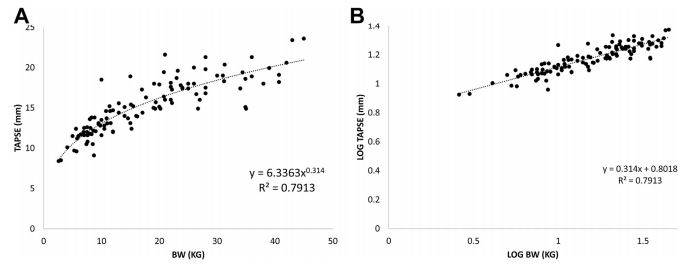
TAPSE, tricuspid annular plane systolic excursion; BW, bodyweight.
Figure 2 The ratio, TAPSE:Ao has a weak negative relationship with bodyweight in both (A) healthy dogs and (B) dogs with mitral valve disease but without pulmonary hypertension
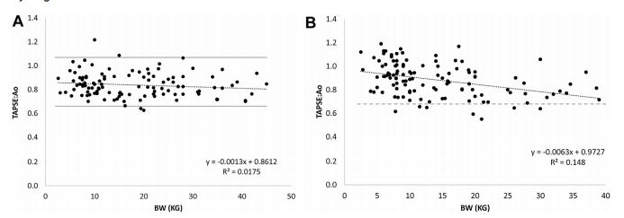
The dashed line represents the lower reference limit forTAPSE:Ao (0.65). TAPSE:Ao, tricuspid annular plane systolic excursion-to-aortic ratio; BW, bodyweight.
Effect of myxomatous mitral valve disease severity on TAPSE:Ao
Effect of pimobendan on TAPSE:Ao in dogs with PH
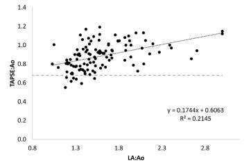
The ratio, TAPSE:Ao, showed a weak positive association with LA:Ao (r2 = 0.2, p < 0.0001; Fig. 3) in dogs with MMVD that did not have PH, with a slope of 0.17 (95%CI: 0.11 to 0.24), and a weak positive association with left ventricular fractional shortening (r2 = 0.1, slope = 0.005, p = 0.001; data not shown), but no association with heart rate (r2 = 0.01, p = 0.4; data not shown).
Effect of PH on TAPSE:Ao
The ratio, TAPSE:Ao, decreased with increasing PH, as determined by tricuspid regurgitation velocity, in Because a recent study demonstrated an increase in TAPSE in dogs administered pimobendan [16], we examined the effect of pimobendan in dogs with PH. We identified 62 dogs with PH where medical records reflected pimobendan use: 27 dogs received pimobendan (25 of these 27 dogs had CHF), 35 dogs did not (5 of these 35 dogs had CHF; doses ranged from 0.21 to 0.35 mg/kg q12hrs). In dogs receiving pimobendan and dogs not receiving pimobendan, TAPSE:Ao decreased with increasing PH severity in an almost parallel manner without an apparent effect of pimobendan (Fig. 5, slopes and intercepts not different; p = 0.78 and p = 0.62 respectively).
Figure 3 The ratio TAPSE:Ao has a weak positive linear relationship with left atrial size in dogs with mitral valve disease but without pulmonary hypertension.
Diagnostic performance of TAPSE:Ao in pre¬dicting severe PH
Using the lower reference limit of 0.65 to differentiate dogs with and without all degrees of PH, we found that as the category of PH severity increased, sensitivity of TAPSE:Ao to detect PH improved without a substantial compromise in specificity (Table 1). The optimal criterion, calculated from the Youden index, ranged from 0.69 to 0.75, slightly higher than the lower reference limit of 0.65. This improved sensitivity for each category of PH, but reduced specificity.
Figure 4 The ratio TAPSE:Ao has a modest negative linear relationship with tricuspid regurgitation velocity in dogs with pulmonary hypertension
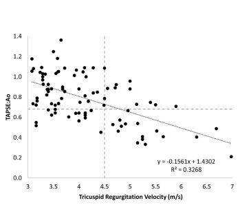
Horizontal dashed line represents the lower reference limit for TAPSE:Ao (0.65). Vertical dashed line represents the lower thresh¬old for severe pulmonary hypertension (pressure gradient = 80 mmHg). TAPSE:Ao: tricuspid annular plane systolic excursion-to-aortic ratio; LA:Ao; left atrial-to-aortic ratio.
Figure 5 The ratio TAPSE:Ao is not affected by administration of pimobendan in dogs with pulmonary hypertension
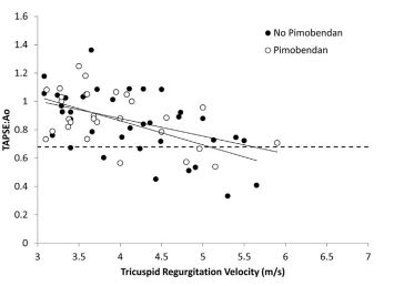
Dogs receiving pimobendan (open circles) and not receiving pimobendan (black circles). TAPSE:Ao, tricuspid annular plane systolic excursion-to-aortic ratio.
In dogs with MMVD and PH, TAPSE:Ao was related to the size of the left atrium, similar to dogs with MMVD but without PH. In the 91 dogs with PH, TAPSE:Ao was <0.65 in 9/64 dogs with LA:Ao > 1.6 (Fig. 6). Three dogs had mild PH, two dogs had moderate PH, and four dogs had severe PH. Of the 15 dogs with severe PH and LA:Ao > 1.6, 4 had TAPSE:Ao < 0.65. However, of the 13 dogs with severe PH and LA:Ao < 1.6,11 had TAPSE:Ao < 0.65.
Diagnostic performance of TAPSE:Ao(adj) in detecting severe PH
To correct for the apparent 'normalizing’ effect of MMVD on TAPSE:Ao, we regressed TAPSE:Ao on LA:Ao in dogs with MMVD but without PH (Fig. 3). Using this equation, we determined the TAPSE:Ao for a dog with an LA:Ao = 1.6 to provide a threshold correction value (0.89). We then adjusted the TAPSE:Ao for dogs with LA:Ao > 1.6 and concurrent PH as follows
TAPSE:AO(adj) = TAPSE:Ao - 0.174 * LA.Ao - 0.61 + 0.89
Regression of TAPSE:Ao on LA:Ao in dogs with MMVD and no pulmonary hypertension
Value of TAPSE:Ao at an LA:Ao of 1.6 in dogs with MMVD and no pulmonary hypertension
Table 1 Diagnostic performance of TAPSE:Ao and TAPSE:Ao(adj) in diagnosis of pulmonary hypertension of varying severity in dogs.
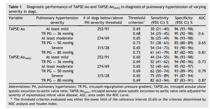
The adjusted ratio, TAPSE:Ao(adj), improved the sensitivity of detecting dogs with severe PH and MMVD with LA enlargement without decreasing specificity (Fig. 7A). Specifically, of the 15 dogs with severe PH and LA:Ao > 1.6, 9 had TAPSE:Ao(adj) < 0.65, consistent with a diagnosis of PH, whereas only 4 of these 15 dogs had TAPSE:Ao < 0.65 (Fig. 7B). The sensitivity of TAP- SE:Ao(adj) for detecting severe PH increased from 54% (for TAPSE:Ao) to 75% and specificity decreased from 93% to 91% (p = 0.007 for com¬parison of the two ROC curves, Table 1). In addition, 9/64 dogs with LA:Ao > 1.6 and PH of various severities had a TAPSE:Ao < 0.65, but this increased to 20/64 dogs when using the TAPSE:Ao(adj).
Figure 6 The ratio TAPSE:Ao in dogs with pulmonary hypertension has a positive relationship with left atrial size
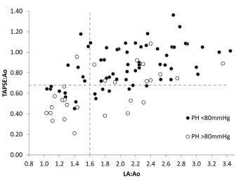
Dogs with severe pulmonary hypertension (open circles) and left atrial enlargement mostly have TAPSE:Ao > 0.65. Horizontal dashed line represents the lower reference limit for TAPSE:Ao (0.65). Vertical dashed line represents the lower threshold for left atrial enlargement (LA:Ao = 1.6). TAPSE:Ao, tricuspid annular plane systolic excursion-to-aortic ratio; LA:Ao, left atrial-to-aortic ratio.
When TAPSE:Ao and TAPSE:Ao(adj) were regressed against tricuspid regurgitation velocity for 52 dogs with PH and moderate-severe MMVD (as defined by LA:Ao > 1.9) or 39 dogs with mild MMVD (LA:Ao < 1.9), TAPSE:Ao for the two groups differed (intercepts for the two regression lines differed, p = 0.02), with dogs that had moderate-severe MMVD and PH showing 'preserved’ RV function across the range of PH severity (Fig. 8A). However, after correcting for LA:Ao (TAP- SE:Ao(adj)), both groups showed similar relationships with tricuspid regurgitation velocity (intercepts and slopes not different, p = 0.4), with 'unmasking’ of decreased RV function in the dogs with PH and more severe MMVD (Fig. 8B). All data for wTAPSE are included in Appendix A. In brief, wTAPSE had a lower limit of the reference interval of 0.59 and showed diagnostic utility similar to TAPSE:Ao in identifying severe PH.
Discussion
Our study shows that normalization of TAPSE by creating a TAPSE:Ao (or wTAPSE) removes the effect of bodyweight and provides a single lower reference limit of 0.65 (or 0.59 for wTAPSE and 0.47 for nTAPSE) that is applicable across dogs of various bodyweights. Our findings suggest that TAPSE:Ao and wTAPSE behave similarly in dogs with MMVD and dogs with PH and findings for wTAPSE (Appendix A) mostly mirror those of TAP- SE:Ao.g Although there is a statistically significant decrease in TAPSE:Ao with bodyweight, this effect is small enough to be clinically irrelevant, because bodyweight predicts less than 2% of the variability in TAPSE:Ao.
Figure 7 (A) TAPSE:AO(adj) (black circles) increases the probability of detecting dogs with pulmonary hypertension and increased left atrial size, compared with TAPSE:Ao (open circles)
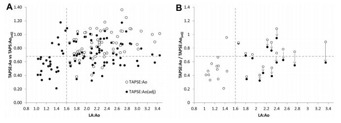
Black circles without corresponding open circles have identical TAPSE:Ao and TAPSE:Ao(adj). (B) Subset of dogs with severe pulmonary hypertension highlighting the decrement in TAPSE:Ao after adjustment for left atrial size (TAPSE:Ao(adj)). A total of 9/15 dogs with severe pulmonary hypertension and left atrial enlargement have TAPSE:Ao(adj) < 0.65; only 4 of these dogs have TAPSE:Ao < 0.65. Horizontal dashed line represents the lower reference limit for TAPSE:Ao (0.65). Vertical dashed line represents the lower threshold for left atrial enlargement (LA:Ao = 1.6). TAPSE:Ao, tricuspid annular plane systolic excursion-to-aortic ratio. TAPSE:Ao(adj), tricuspid annular plane systolic excursion-to-aortic ratio adjusted for severity of mitral valve disease. LA:Ao, left atrial-to-aortic ratio.
In dogs with MMVD and no PH, TAP- SE:Ao shows a similar relationship to bodyweight. However, in these dogs with MMVD but without PH, TAPSE:Ao increases with increasing severity of MMVD—a heretofore unrecognized phenomenon. In dogs with MMVD and PH TAPSE:Ao is unaffected by co-administration of pimobendan. Importantly, TAPSE:Ao decreases linearly with increasing pul¬monary artery pressure. Owing to the common association of MMVD and PH [5,17], TAPSE:Ao provides a specific, but insensitive method for diagnosing PH in dogs with MMVD, because of the 'preservation’ of TAPSE:Ao with more severe forms of MMVD. However, adjusting TAPSE:Ao for the degree of LA enlargement modestly improves the sensitivity for diagnosing severe PH.
g Because TAPSE:Ao and wTAPSE behave similarly, we have limited the discussion to TAPSE:Ao, which most readers will be likely to use, but wTAPSE data can be viewed in Appendix A and specifics regarding wTAPSE will be highlighted in instances where wTAPSE differed substantially from TAPSE:Ao.
Our findings are similar to, and extend on, the previous study of Pariaut et al. [1]. Indeed, part of the data set in the current study comprised of dogs used in that previous study. The larger cohorts of healthy dogs and dogs with PH strengthen the initial observations. Our study supports the initial observations of Pariaut et al that TAPSE:Ao (like TAPSE) has poor sensitivity, but good specificity in diagnosing severe PH—in other words, a normal TAPSE:Ao does not exclude the possibility of PH, but a lowTAPSE:Ao is strongly supportive of the diagnosis. In addition, our investigation of TAPSE:Ao is not unique—Pariaut et al. noted the curvilinear relationship of TAPSE and bodyweight but elected not to examine a normalized ratio in their study because they were concerned about the variability associated with aortic valve measurements. However, other studies have shown that this variability is relatively small, provided a consistent diastolic time-point is used, thereby supporting the idea of TAPSE:Ao as a reasonable bodyweight-independent estimate of right ventricular systolic function [12,18].
Our results differ from those of other investigators who examined the effect of MMVD on TAPSE. Tidholm et al. [4] found that, in the pres¬ence of MMVD, nTAPSE did not accurately predict the presence of PH. This might be explained in part by our observation that TAPSE:Ao is 'preserved’ with worsening MMVD (as defined by an increasing LA:Ao), which essentially masks the effect of PH on TAPSE:Ao, wTAPSE and, presumably, nTAPSE (which is qualitatively identical to wTAPSE). Unlike our study, those investigators did not compare the effect of PH on TAPSE in dogs without MMVD. Poser et al. [5] found an even more perplexing result—that nTAPSE did not change with worsening stages of MMVD or worsening PH. Indeed, examination of the plots in that study suggests that nTAPSE trended upward, rather than downward, with worsening PH in the presence of MMVD. Again, these findings might reflect both the 'masking’ effect of MMVD on TAPSE, and the combining, in both of these studies, of multiple categories of PH (i.e., moderate and severe PH was grouped together in both studies). Given our observation (and that of Pariaut et al. [1]) that tricuspid annular plane systolic excursion (TAPSE) and TAPSE:Ao (and wTAPSE) are specific but insensitive methods of identifying severe PH, it is not surprising that previous studies which combined less severe grades of PH with more severe grades failed to observe a difference in nTAPSE between groups.
Figure 8 (A) TAPSE:Ao in dogs with pulmonary hypertension and more advanced myxomatous mitral valve disease (open circles) and in dogs with pulmonary hypertension and mild or absent myxomatous mitral valve disease (black circles) have different relationships with tricuspid regurgitation.
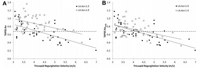
(B) TAPSE:Ao(adj) in dogs with pulmonary hyper¬tension and more severe myxomatous mitral valve disease (open circles) corrects the relationship with tricuspid regurgitation, allowing for increased detection of pulmonary hypertension in these dogs. TAPSE:Ao: tricuspid annular plane systolic excursion-to-aortic ratio; TAPSE:Ao(adj), tricuspid annular plane systolic excursion-to-aortic ratio adjusted for severity of mitral valve disease.
We evaluated the impact of MMVD without PH on TAPSE:Ao. Poser et al. [5] also examined nTAPSE in dogs with MMVD but did not present data for these dogs to enable direct comparisons with our findings. The dogs in our study all had MMVD of varying severity, ranging from mild to severe. Our data suggest that TAPSE:Ao increases with increasingly severe MMVD. For this reason, the depressant effect of PH on TAP- SE:Ao (wTAPSE, nTAPSE, and TAPSE) might be dimin¬ished in dogs with MMVD. Indeed, 55/64 dogs with PH and left atrial enlargement secondary to MMVD had TAPSE:Ao > 0.65 and 56/64 dogs had wTAPSE >0.59.
Notably, 11/15 dogs with severe PH and left atrial enlargement had TAPSE:Ao > 0.65 and 10/15 had wTAPSE > 0.59. On the other hand, 11/13 dogs with severe PH and no evidence of left heart disease (normal left atrial size) had TAPSE:Ao < <0.65 and wTAPSE < 0.59. The mechanism by which tricuspid annular plane systolic excursion (TAPSE) increases with worsening mitral valve disease cannot be determined in this study. A plausible hypothesis is thatchangesin loading conditions of the left ventricle in dogs with MMVD alters systolic translational forces on the right heart, resulting in an increasing TAP- SE:Ao, as suggested by Poser et al [5]. Indeed, TAP- SE:Ao was very weakly correlated with left ventricular fractional shortening, consistent with this hypothesis (data not shown).
Recently, investigators demonstrated an increase in tricuspid annular plane systolic excursion (TAPSE) in healthy dogs after a single dose of pimobendan [16]. However, we could find no effect on TAPSE:Ao in dogs with PH that were administered pimobendan. Our findings suggest that once the right ventricle is subjected to increased afterload, the effect of pimobendan on longitudinal ventricular motion might be marginal.
In light of the 'masking’ effect of MMVD and potentially drug therapy on TAPSE:Ao, we adjusted for the effect by normalizing to an upper limit of LA:Ao of 1.6 by incorporating the regression equation for TAPSE:Ao (or wTAPSE) on LA:Ao derived from dogs with MMVD but no PH. The TAPSE:Ao(adj) improved the sensitivity of detecting severe PH in dogs with concurrent more MMVD and left atrial enlargement, without affecting the detection of dogs with PH in dogs that had either no evidence of MMVD, or mild MMVD, with normal left atria. The adjustment did not alter the spe¬cificity of detection (it did not increase the false positive rate), although the number of dogs with severe PH and moderate-severe MMVD in our study was small (14 dogs). Clinicians could potentially incorporate this correction for TAPSE:Ao into the software packages of ultrasound machines to automatically calculate both TAPSE:Ao and TAP- SE:Ao(adj), and then use TAPSE:Ao if the LA:Ao < 1.6 and TAPSE:Ao(adj) if the LA:Ao > 1.6.
Although we considered the effect of bodyweight on TAPSE:Ao to be clinically unimportant, we did observe a very slight negative association between these two variables. This might be explained by the different scaling exponents for TAPSE and for Ao, when regressed against bodyweight—for all three observers, scaling exponents for Ao were marginally higher than for tricuspid annular plane systolic excursion (TAPSE). This could create a very slight negative slope, as we observed.
Thirty-nine of our dogs had convincing evidence of precapillary PH (no evidence of left heart disease or only mild MMVD). Of these 39 dogs, 16 had severe PH; 3 had mild LA enlargement (1.6 < LA:Ao < 1.9) and 13 had LA:Ao < 1.6. In the 2/3 dogs with mild LA enlargement and severe PH, TAPSE:Ao was >0.65, supporting the idea of a 'masking’ effect of MMVD on TAPSE:Ao. However, TAPSE:Ao(adj) decreased to 0.69 in one of these dogs, suggesting that even with suspected severe precapillary PH and concomitant MMVD, TAPSE:Ao(adj) can improve detection of severe PH. Therefore, the diagnostic utility of unadjusted TAPSE:Ao (and TAPSE) in diagnosis of precapillary, rather than post-capillary, PH, should be reassessed with larger patient populations (although TAPSE:Ao(adj) would work equally well, since these dogs mostly have no correction applied by the adjustment formula, as they have LA:Ao < 1.6). In addition, in cases with precapillary PH and concomitant MMVD, TAPSE:Ao(adj) might improve identification of severe PH.
The investigators all had similar ranges of tricuspid annular plane systolic excursion (TAPSE) and Ao measurements for healthy dogs, and dogs with MMVD and no PH, consistent with the inter¬observer variability reported by Pariaut et al. [1]. Therefore, provided correct imaging planes are obtained, our data should be applicable to other studies of TAPSE:Ao. Clinicians should recognize that TAPSE, TAP- SE:Ao, and TAPSE:Ao(adj) are all likely to be decreased in dogs with primary right ventricular systolic dysfunction, and that these measurements are not exclusively affected by PH. However, the most common application of TAPSE has been in assessing right ventricular longitudinal function in the setting of PH. Finally, we took the opportunity to provide a lower reference limit for nTAPSE, which has been investigated by previous investigators [4,5], but without establishing reference limits.
Our study provides clinicians with a single lower reference limit of 0.65 for TAPSE:Ao, 0.59 for wTAPSE, and 0.47 for nTAPSE, that can be applied to all dogs. This should simplify the use of tricuspid annular plane systolic excursion (TAPSE) and encourage cardiologists to measure TAPSE:Ao, wTAPSE, or nTAPSE in dogs with suspected severe PH which cannot be confirmed by spectral Doppler echocardiography and in which other causes of right ventricular systolic dysfunction have been excluded. Furthermore, in dogs with concomitant LA enlargement secondary to MMVD, a mathematical correction of TAPSE:Ao can improve detection of severe PH.
Appendix A
Results. Relationship between wTAPSE or nTAPSE and bodyweight
Weight-adjusted TAPSE, wTAPSE, showed no rela¬tionship with bodyweight in both healthy dogs (r2 = 0.03, p = 0.07; Figure A1), and a very weak negative relationship with bodyweight in dogs with MMVD but without pulmonary hypertension (PH; r2 = 0.05, p < 0.04; Figure A2), with a slope of -0.0015 (95%CI: -0.002 to -0.0004) and -0.004 (95%CI: -0.007 to -0.0001), respectively. The ref¬erence intervals extended from 0.59 (95%CI: 0.56 to 0.66) to 0.97 (95%CI: 0.87 to 1.08). Using the lower reference limit of 0.59, only 3/137 healthy dogs and 1/115 dogs with MMVD (all of which had no left atrial enlargement) had values below this limit.
Figure A1 The index, wTAPSE has a very weak negative relationship with bodyweight
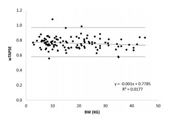
The gray lines rep¬resent the lower and upper reference limits for wTAPSE (0.59 and 1.07). wTAPSE(adj), weight-adjusted tricuspid annular plane systolic excursion-to-aortic valve ratio corrected for mitral valve disease severity.
Figure A2 The index, wTAPSE in dogs with mitral valve disease but without pulmonary hypertension has a very weak negative relationship with bodyweight, with most dogs having wTAPSE > 0.59
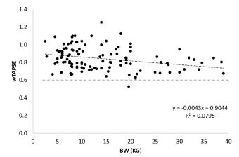
wTAPSE(adj), weight-adjus¬ted tricuspid annular plane systolic excursion-to-aortic valve ratio corrected for mitral valve disease severity.
Normalized TAPSE, nTAPSE, also showed no rela¬tionship with bodyweight (r2 = 0.025, p = 0.07), with a slope of -0.0011 (95%CI: -0.002 to -0.0001). The reference intervals extended from 0.47 (95%CI: 0.45 to 0.53) to 0.78 (95%CI: 0.70 to 0.87). Using the lower reference limit of 0.47, only 2/137 healthy dogs had values below this limit.
Effect of myxomatous mitral valve disease severity on wTAPSE
Weight-adjusted TAPSE, wTAPSE, showed a pos¬itive association with LA:Ao (r2 = 0.07, P = 0.005; Figure A3), with a slope of 0.1 (95%CI: 0.03 to 0.16).
Figure A3 The index, wTAPSE has a weak positive linear relationship with left atrial size in dogs with mitral valve disease but without pulmonary hypertension
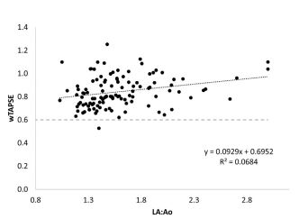
wTAPSE(adj), weight-adjusted tricuspid annular plane systolic excursion-to-aortic valve ratio corrected for mitral valve disease severity.
Effect of PH on wTAPSE
Weight-adjusted TAPSE, wTAPSE, decreased with increasing PH, as determined by tricuspid regur¬gitation velocity, in a linear manner (r2 = 0.3, p < 0.0001; Figure A4), with a slope of -0.14 (95% CI: -0.17 to -0.08).
Figure A4 The index, wTAPSE has a modest negative linear relationship with tricuspid regurgitation velocity in dogs with pulmonary hypertension. Horizontal dashed line represents the lower reference limit for wTAPSE (0.59).
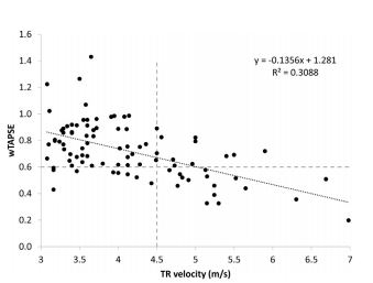
Vertical dashed line represents the lower threshold for severe pulmonary hypertension (pressure gradient = 80 mmHg). wTAPSE(adj), weight-adjusted tricuspid annular plane systolic excursion-to-aortic valve ratio corrected for mitral valve disease severity.
Diagnostic performance of wTAPSE in predicting severe PH
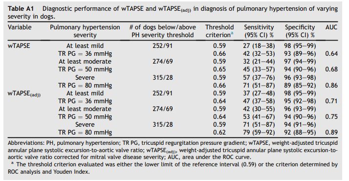
Using the lower reference limit of 0.59 to differentiate dogs with and without all degrees of PH, wTAPSE showed increasing sensitivity without a substantial compromise in specificity in identifying increasingly more severe categories of PH (Table A1). The optimal criteria, calculated from the Youden index was 0.66, slightly higher than the lower ref¬erence limit of 0.59.
In dogs with PH, wTAPSE was also related to the size of the left atrium. Inthe91 dogs with PH, wTAPSE was <0.59 in 8/64 dogs with LA:Ao > 1.6 (Figure A5). Of the 15 dogs with severe PH and LA:Ao > 1.6, 5 had wTAPSE < 0.59. However, of the 13 dogs with severe PH and LA:Ao < 1.6, 11 had wTAPSE < 0.59.
Diagnostic performance of wTAPSE(adj) in detecting severe PH
To correct for the apparent 'normalizing’ effect of mitral valve disease on wTAPSE, we regressed wTAPSE on LA:Ao in dogs without PH (Figure A5).
Table A1 Diagnostic performance of wTAPSE and wTAPSE(adj) in diagnosis of pulmonary hypertension of varying severity in dogs
Using this equation, we determined the wTAPSE for a dog with an LA:Ao = 1.6 to provide a threshold correction value (0.84). We then adjusted the wTAPSE for dogs with LA:Ao > 1.6 and concurrent PH as follows:
TAPSE:Ao(aai) = TAPSE: Ao - 0.174 *LA:Ao- 0.61 + 0.89
Regression of TAPSE:Ao on LA:Ao in dogs with MMVD and no pulmonary hypertension
Value of TAPSE:Ao at an LA:Ao of 1.6 in dogs with MMVD and no pulmonary hypertension
wTAPSE that was corrected for mitral valve disease, wTAPSE(adj), improved the sensitivity of detecting dogs with severe PH and MMVD without decreasing specificity (Figure A6). Specifically, of the 15 dogs with severe PH and LA:Ao > 1.6, 9 had wTAPSE(adj) < 0.59, consistent with a diagnosis of PH, whereas only 5 of these 15 dogs had wTAPSE < 0.59 (Fig. 7B). The sensitivity of wTAP- SE(adj) for detecting severe PH increased from 57% (for wTAPSE) to 71% and specificity decreased from 96% to 94% (p = 0.01 for comparison of the two ROC curves, Table A1).
In addition, 8/64 dogs with LA:Ao > 1.6 and PH of various severities had a wTAPSE < 0.59, but this increased to 17/64 dogs when using the wTAPSE(adj).
Figure A5 The index, wTAPSE in dogs with pulmonary hypertension has a positive relationship with left atrial size. wTAPSE(adj) (black circles) increases the probability of detecting dogs with pulmonary hypertension and increased left atrial size, compared with wTAPSE (open circles).
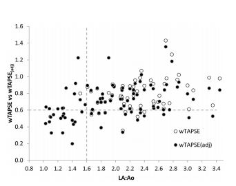
Black circles without corresponding open circles have identical wTAPSE and wTAPSE(adj). Horizontal dashed line represents the lower reference limit for wTAPSE (0.59). Vertical dashed line represents the lower threshold for left atrial enlargement (LA:Ao = 1.6). wTAPSE(adj), weight-adjusted tricuspid annular plane systolic excursion-to-aortic valve ratio corrected for mitral valve disease severity; LA:Ao, left atrial-to-aortic ratio.
Figure A6 Subset of dogs with severe pulmonary hypertension highlighting the decrement in wTAPSE after adjustment for left atrial size (wTAPSE(adj)).
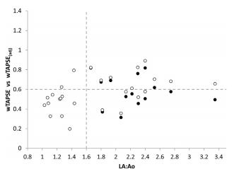
A total of 9/15 dogs with severe pulmonary hypertension and left atrial enlargement have wTAPSE(adj) < 0.59, whereas only 5 of these dogs have wTAPSE < 0.59. Horizontal dashed line represents the lower reference limit for wTAPSE (0.59). Vertical dashed line represents the lower threshold for left atrial enlargement (LA:Ao = 1.6). wTAPSE(adj), weight-adjusted tricuspid annular plane systolic excursion-to-aortic valve ratio corrected for mitral valve disease severity; LA:Ao, left atrial-to-aortic ratio.
References
- Pariaut R, Saelinger C, Strickland KN, Beaufrere H, Reynolds CA, Vila J. Tricuspid annular plane systolic excursion (TAPSE) in dogs: reference values and impact of pulmonary hypertension. J Vet Intern Med 2012;26: 1148-54.
- Visser LC, Scansen BA, Schober KE, Bonagura JD. Echo- cardiographic assessment of right ventricular systolic function in conscious healthy dogs: repeatability and ref¬erence intervals. J Vet Cardiol 2015;17:83-96.
- Kaye BM, Borgeat K, Motskula PF, Luis Fuentes V, Connolly DJ. Association of tricuspid annular plane systolic excursion with survival time in boxer dogs with ventricular arrhythmias. J Vet Intern Med 2015;29:582-8.
- Tidholm A, Hoglund K, Haggstrom J, Ljungvall I. Diagnostic value of selected echocardiographic variables to identify pulmonary hypertension in dogs with myxomatous mitral valve disease. J Vet Intern Med 2015;29:1510-7.
- Poser H, Berlanda M, Monacolli M, Contiero B, Coltro A, Guglielmini C. Tricuspid annular plane systolic excursion in dogs with myxomatous mitral valve disease with and without pulmonary hypertension. J Vet Cardiol 2017;19: 228-39.
- Visser LC, Sloan CQ, Stern JA. Echocardiographic assessment of right ventricular size and function in cats with hypertrophic cardiomyopathy. J Vet Intern Med 2017;31: 668-77.
- Spalla I, Payne JR, Borgeat K, Pope A, Fuentes VL, Connolly DJ. Mitral annular plane systolic excursion and tricuspid annular plane systolic excursion in cats with hypertrophic cardiomyopathy. J Vet Intern Med 2017;31: 691-9.
- Mertens LL, Friedberg MK. Imaging the right ventricle-current state of the art. Nat Rev Cardiol 2010;7: 551 -63.
- Lopez-Candales A, Dohi K, Rajagopalan N, Edelman K, Gulyasy B, Bazaz R. Defining normal variables of right ventricular size and function in pulmonary hypertension: an echocardiographic study. Postgrad Med J 2008;84:40-5.
- Koestenberger M, Ravekes W, Everett AD, Stueger HP, Heinzl B, Gamillscheg A, et al. Right ventricular function in infants, children and adolescents: reference values of the tricuspid annular plane systolic excursion (TAPSE) in 640 healthy patients and calculation of z score values. J Am Soc Echocardiogr 2009;22:715-9.
- Brown DJ, Rush JE, MacGregor J, Ross Jr JN, Brewer B, Rand WM. M-mode echocardiographic ratio indices in normal dogs, cats, and horses: a novel quantitative method. J Vet Intern Med 2003;17:653-62.
- Dickson D, Caivano D, Patteson M, Rishniw M. The times they are a-changin': two-dimensional aortic valve measurements differ throughout diastole. J Vet Cardiol 2016; 18:15-25.
- Rishniw M, Erb HN. Evaluation of four 2-dimensional echocardiographic methods of assessing left atrial size in dogs. J Vet Intern Med 2000;14:429-35.
- Cornell CC, Kittleson MD, Della Torre P, Haggstrom J, Lombard CW, Pedersen HD, et al. Allometric scaling of M- mode cardiac measurements in normal adult dogs. J Vet Intern Med 2004;18:311-21.
- Geffre A, Concordet D, Braun J-P, Trumel C. Reference Value Advisor: a new freeware set of macroinstructions to calculate reference intervals with Microsoft Excel. Vet Clin Pathol 2011;40:107-12.
- Visser LC, Scansen BA, Brown NV, Schober KE, Bonagura JD. Echocardiographic assessment of right ven¬tricular systolic function in conscious healthy dogs fol¬lowing a single dose of pimobendan versus atenolol. J Vet Cardiol 2015;17:161-72.
- Serres FJ, Chetboul V, Tissier R, Carlos Sampedrano C, Gouni V, Nicolle AP, et al. Doppler echocardiography- derived evidence of pulmonary arterial hypertension in dogs with degenerative mitral valve disease: 86 cases (2001-2005). J Am Vet Med Assoc 2006;229:1772-8.
- Georgiev R, Rishniw M, Ljungvall I, Summerfield N. Common two-dimensional echocardiographic estimates of aortic linear dimensions are interchangeable. J Vet Cardiol 2013;15:131-8.
^Наверх


